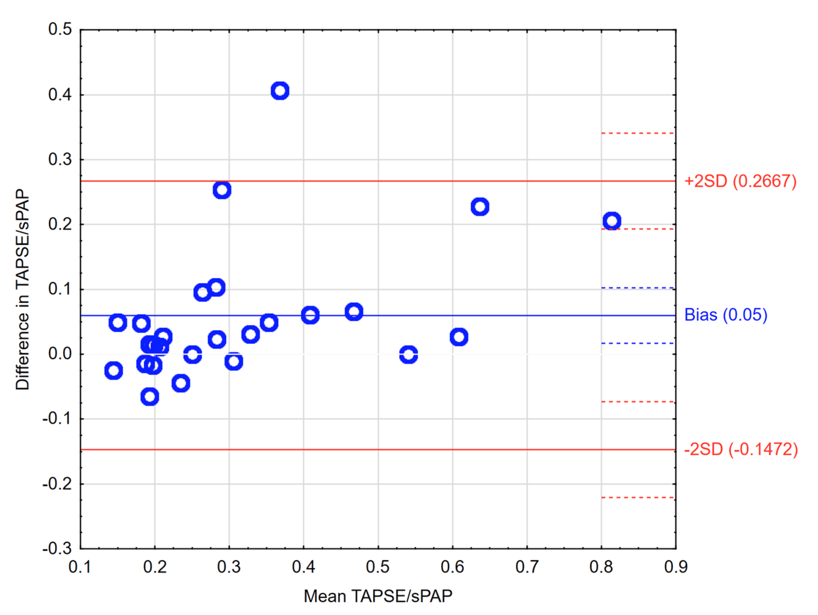
Using echocardiography, researchers were able to examine the association between short and longterm mortality with increasing pulmonary pressures Researchers evaluated eRVSP levels for 74,405 men and ,437 women who were, on average, 656 years of ageThe CE was significantly more frequent in 19 (92%) patients with TRPG/TAPSE >45 than in 1 (908%) with TRPG/TAPSE ≤45 (4 (211%) vs 4 (21%), P=) Among normotensive APE A value of > 130 ms is normal, while < 100 ms is highly suggestive of pulmonary hypertension Mean pulmonary pressure is calculated by the formula mPAP = 90 − (062*AT RVOT) For example, if the AT RVOT is 80 ms, the mPAP = 90 −(062*80), that is 404 mmHg (normal < 25 mmHg)

Prognostic Significance Of Right Ventricular Function During Exercise In Asymptomatic Minimally Symptomatic Patients With Nonobstructive Hypertrophic Cardiomyopathy Hirasawa Echocardiography Wiley Online Library
What does a normal echo mean
What does a normal echo mean-The subcostal view is obtained with the patient in the supine position Position of the transducer The transducer is placed, with the index marker pointing towards the patient's left breast, 3 cm below the xiphoid process The transducer should have an angle of incidence of approximately 45° towards the thorax So you've been referred by your doctor or other health care provider to have an echocardiogram, or ultrasound of your heart If you've had an echocardiogram done in the past, then you pretty much know what to expect But if this is your first time having an echocardiogram, then you've probably got a few questions That's ok You're not alone


Characteristics And Prognostic Associations Of Echocardiographic Pulmonary Hypertension With Normal Left Ventricular Systolic Function In Patients 90 Years Of Age American Journal Of Cardiology
Yet the initial TPG was, on average, >12 mmHg, with a range of values from 6– mmHg Since transplantation did not always normalise mean Ppa, the data taken from 12 out of the patients in whom mean Ppa decreased to below the value of 25 mmHg were reexamined The preoperative TPG was >12 mmHg (range 12– mmHg) in six of the patients E/e' The normal E/e' ratio from the medial annulus is 15 is indicative of an elevated left atrial pressure or PCWP (>18mmHg) The ranges for E/e' from the lateral mitral annulus are 10 respectively 35A transesophageal echocardiogram (TEE) uses echocardiography to assess how well the heart works During the procedure, a transducer (like a microphone) sends out ultrasonic sound waves at a frequency too high to be heard When the transducer is placed at certain locations and angles, the ultrasonic sound waves move through the skin and other body tissues to the heart tissues, where
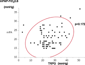
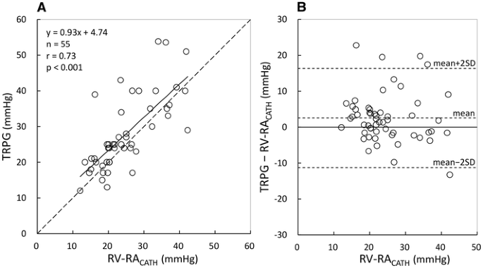
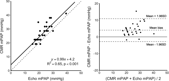
Normal Echo Values Below is a complete and thorough list of normal echo values This list of normal echo values is from echopediaorg Left Ventricle Left Ventricular Systolic Function Reference limits and values and partition values of left ventricular functionASE Women Men Reference range Mildly abnormal Moderately abnormal Severely abnormalThe frequency and proportion of patients in each diagnos tic category are summarized in Table 3Amongthepa tients who had increased CMR mPAP but normal TRPG by echocardiography, TRPG was 27 –30 mmHg Comparison between estimated mPAP by CMR 4D flow analysis and TRPG by echocardiography by Dopplerestimated RVSP has varied between 30 and 45 mmHg in different studies1,5,12,13The cutoff value for RVSP to diagnose clinically relevant PH has varied between 45 and 56 mmHg1,13A TRPG >55 mmHg is a
Based on echocardiographic TRPG measurements, patients with IPF were classified into two groups named high TRPG (TRPG ≥30 mmHg) and normal TRPG (TRGP Echocardiogram results showed normal ej fx of 6065%, mild pulmonary htn, tricuspid regurgatation with right ventricle measuring 3639 by Doppler everything else on Doppler was normal EKG, chest Xray and stress test normalA TTE is a procedure used to check for problems with your heart It will also show any problems in the blood vessels near your heart Sound waves are sent through a handheld device placed on your chest The sound waves show the structure and function of your heart through pictures on a monitor


What Is Normal Mean Pulmonary Arterial Pressure


Tricuspid Regurgitation Peak Gradient Trpg Tricuspid Annulus Plane Systolic Excursion Tapse A Novel Parameter For Stepwise Echocardiographic Risk Stratification In Normotensive Patients With Acute Pulmonary Embolism Semantic Scholar
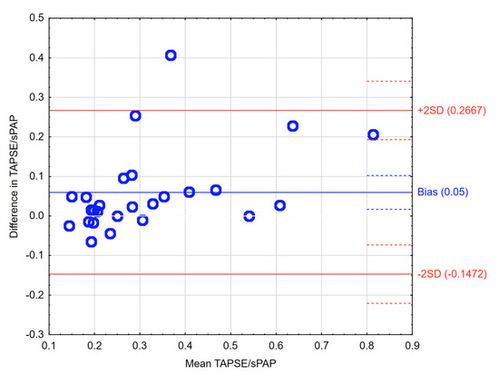
Pulmonary hypertension (PH) due to left heart disease, classified as group 2 according to the Dana Point 08 classification, is believed to be the most common cause of PH and is associated with high morbidity and mortality Epidemiological studies of group 2 PH are less exhaustive than for rarer causes of PH such as isolated pulmonaryThe tricuspid annulus plane systolic excursion (TAPSE) and tricuspid regurgitation peak gradient (TRPG) can be used for risk stratification, so we analyzed the prognostic value of a new echo parameter (TRPG/TAPSE) for prediction of APErelated 30day death or need for rescue thrombolysis in initially normotensive APE patientsMethods and ResultsThe study group consists of 400 nonhighrisk APE patients (191 men, age 631±1 years) who had undergone echocardiographyIt is normal for your heart rate, blood pressure, breathing rate and perspiration to increaseAs you stop exercising suddenly, it is normal to feel a little unsteady when getting off the treadmill and onto the exam table for the echocardiogram How long does the test take?


14 Guidelines Of Taiwan Society Of Cardiology Tsoc For The Management Of Pulmonary Arterial Hypertension Abstract Europe Pmc
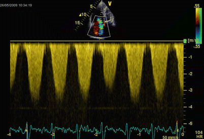


Advanced Imaging In Pulmonary Hypertension Thoracic Key
The appointment will take about 60 minutes The resulting image of an echocardiogram can show a big picture image of heart health, function, and strength For example, the test can show if the heart is enlarged or has thickened walls Walls thicker than 15cm are considered abnormal They may indicate high blood pressure and weak or damaged valves13 Normal Reference Values for 2DE 6 14 Normal Reference Values for 3DE 6 Recommendation 6 2 LV Global Systolic Function 6 21 Fractional Shortening 6 22 EF 7 23 Global Longitudinal Strain (GLS) 7 24 Normal Reference Values 7 Recommendations 10 3 LV Regional Function 10 31 Segmentation of the Left Ventricle 10 32 Visual


6 Tips For Calculating Rvsp


Echo Features Of Acute Sultania Cardiovascular Sciences Facebook
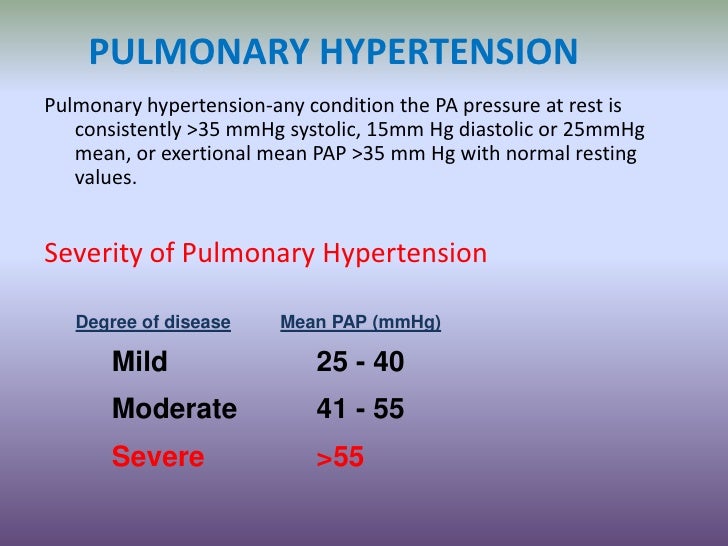
Echo calculates the right ventricular systolic pressure (RVSP) The definition of PH is a pathophysiological condition determined when there is an increase of the MEAN pulmonary artery pressure is > 25 mmHg at rest, obtained by invasive right heart catheterization Unfortunately noninvasive echo obtained values got left out of the definition The echocardiography stress test is very reliable Your doctor will explain your test results to you If the results are normal, your heart is working properly andUsed for risk stratification, so we analyzed the prognostic value of a new echo parameter (TRPG/TAPSE) for prediction of APErelated 30day death or need for rescue thrombolysis in initially normotensive APE patients was normal and/or had normal cardiac biomarkers levels formed the intermediatelowrisk group4


Emdocs Net Emergency Medicine Educationus Probe Tapse Emdocs Net Emergency Medicine Education


Diagnostic Value Of Right Pulmonary Artery Distensibility Index In Dogs With Pulmonary Hypertension Comparison With Doppler Echocardiographic Estimates Of Pulmonary Arterial Pressure Visser 16 Journal Of Veterinary Internal Medicine Wiley
Echocardiography — Once PH is suspected, the initial test of choice is transthoracic echocardiography (TTE), which then determines the sequence of subsequent testing Importantly, patients with evidence of severe PH and/or patients with significant symptoms should be referred to a PH center for rapid evaluation A normal V/Q scanA peak GLS of % can be considered normal The smaller the absolute number, the more abnormal is the strain LV Regional Function Segmentation of the LV Segmentation schemes should reflect coronary perfusion territories and allow standardized communication within echocardiography and with other imaging modalitiesNary hemorrhage45,46 Reported upper normal limits of Dopplerderived SPAP during exercise are


1


1
The mean postexercise TRPG was 403 ± 41 mm Hg (range 1770) and the mean postexercise increase in TRPG was 129 ± 85 mm Hg (range 238) A TRPG increase of 〉 mm Hg was found in 11 (16%) patients (including 4 patients with resting values exceeding 31 mm Hg and 7 patients with normal resting values) normal PAPm at rest is 143 mmHg with an upper limit of normal of approximately mmHg (975th percentile) (Simmonaeu et al, 19) Transthoracic echocardiogram (TTE) remains the most important noninvasive screening tool for PHOrdering an Echo in 16 and Beyond Ask yourself, how is this going to change the management of the patient CCN 15 use echocardiography if, and only if, results have the potential to influence clinical decisions and patient management Responsible utilization


6 Tips For Calculating Rvsp


Characteristics And Prognostic Associations Of Echocardiographic Pulmonary Hypertension With Normal Left Ventricular Systolic Function In Patients 90 Years Of Age American Journal Of Cardiology
The normal value of TAPSE is 24±4 mm Values less than 16 mm indicate impaired systolic function of the right ventricle 5,16 Forfia et al in a prospective study of 63 patients diagnosed with PH showed that the value of TAPSE less than 18 mm was an independent risk factor for mortality in this population 24Learn the how and the why of stroke volume determination with echocardiography In 10 minutesTransthoracic echocardiogram This is the standard test It's like an Xray but without the radiation Specialists use the same technology to check a baby's health before birth


2


Simple Two Dimensional Echocardiographic Scoring System For The Estimation Of Left Ventricular Filling Pressure Journal Of The American Society Of Echocardiography
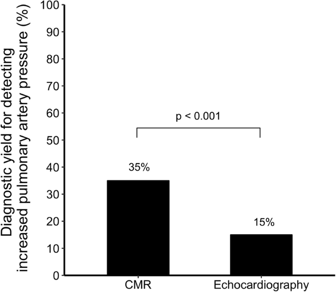
Resting TRPG >31 mmHg and/or positive EDE was found in 30 patients and they were referred to RHC Finally, RHC was performed in patients (16 pts resting TRPG >31 mmHg and 4 others normal resting TRPG and positive EDE) In 12 (60 %) of them an EPAPR with elevated pulmonary capillary wedge pressure (PCWP) was observed Normal values in the neonatal population can be obtained from Koestenberger et al 42 Diminished TAPSE ( Among the patients who had increased CMR mPAP but normal TRPG by echocardiography, TRPG was 27 –30 mmHg Table 3 Frequency of patients in each diagnostic group when comparing cardiovascular magnetic resonance (CMR) and echocardiography (Echo) TR denotes tricuspid regurgitant jet velocity


Pdf Accuracy Of Doppler Echocardiographic Estimates Of Pulmonary Artery Pressures In A Canine Model Of Pulmonary Hypertension


Echocardiography Monitoring Of Pulmonary Hypertension After Pediatric Hematopoietic Stem Cell Transplantation Pediatric Pulmonary Arterial Hypertension And Pulmonary Veno Occlusive Disease After Hematopoietic Stem Cell Transplantation
In case of a normal MV structure, normal LV and LA size, a small central jet area ofNormal >10 mmHg/s (< 27 msec) Boarderline mmHg/s (2732 msec) Abnormal < 1000 mmHg/s (> 32 msec) However, the subclinical high TRPG patients had a higher MELD score than the low/normalTRPG patients (p = 0033), indicating a more severe condition of cirrhosis TTE revealed right ventricular dilatation more frequently in subclinical high TRPG patients than in other patients ( p = 0022)


Diagnostic Value Of Right Pulmonary Artery Distensibility Index In Dogs With Pulmonary Hypertension Comparison With Doppler Echocardiographic Estimates Of Pulmonary Arterial Pressure Topic Of Research Paper In Veterinary Science Download Scholarly
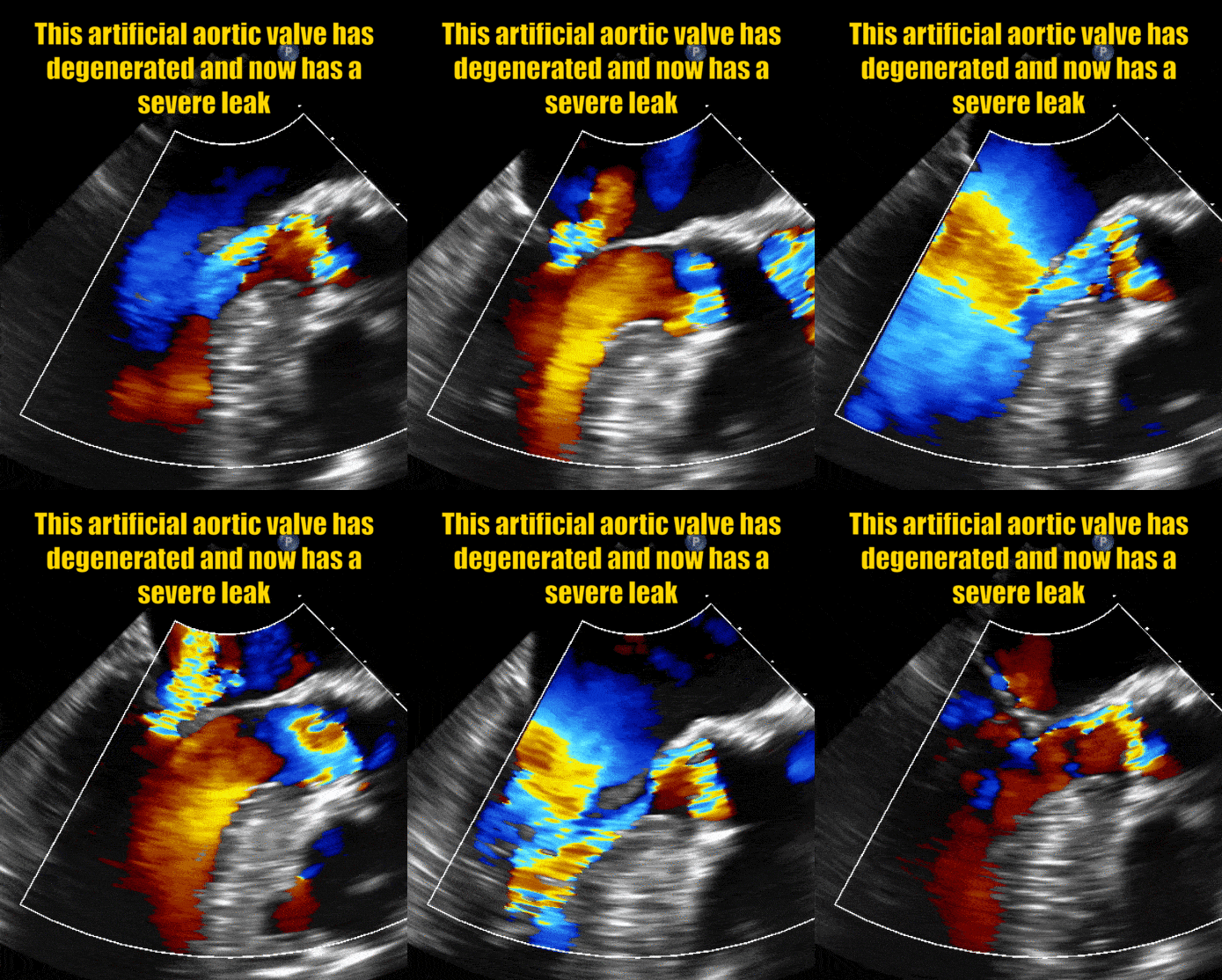


Aortic Regurgitation A Leaky Aortic Valve Myheart
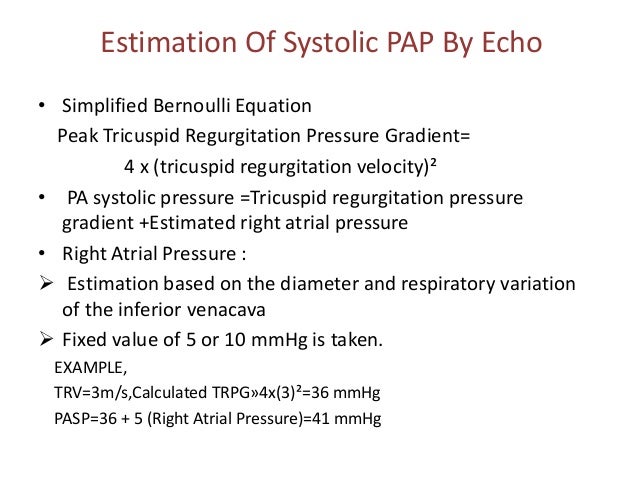
transthoracic echocardiogram Pulmonary arterial hypertension was identified if a tricuspid regurgitation pressure gradient (TRPG) ≥36 mmHg based on echoDoppler assessment was observed–22 The TRPG was derived from the peak systolic tricuspid regurgitation jet velocity using the simplified Bernoulli equation (pressure gradient = 4 mul Although RHC is the only test available to diagnose PH, echocardiography is the preferred screening test for at‐risk patients 6 Current guidelines recommend further evaluation of patients for PH if the tricuspid regurgitation velocity is >29 m/s (corresponding to PASP >≈40 mm Hg) 6 However, our findings demonstrate that PA pressureAn echocardiogram (often called an "echo") is a graphic outline of your heart's movement During this test, highfrequency sound waves, called ultrasound, provide pictures of your heart's valves


Jvs Journal Of Veterinary Science
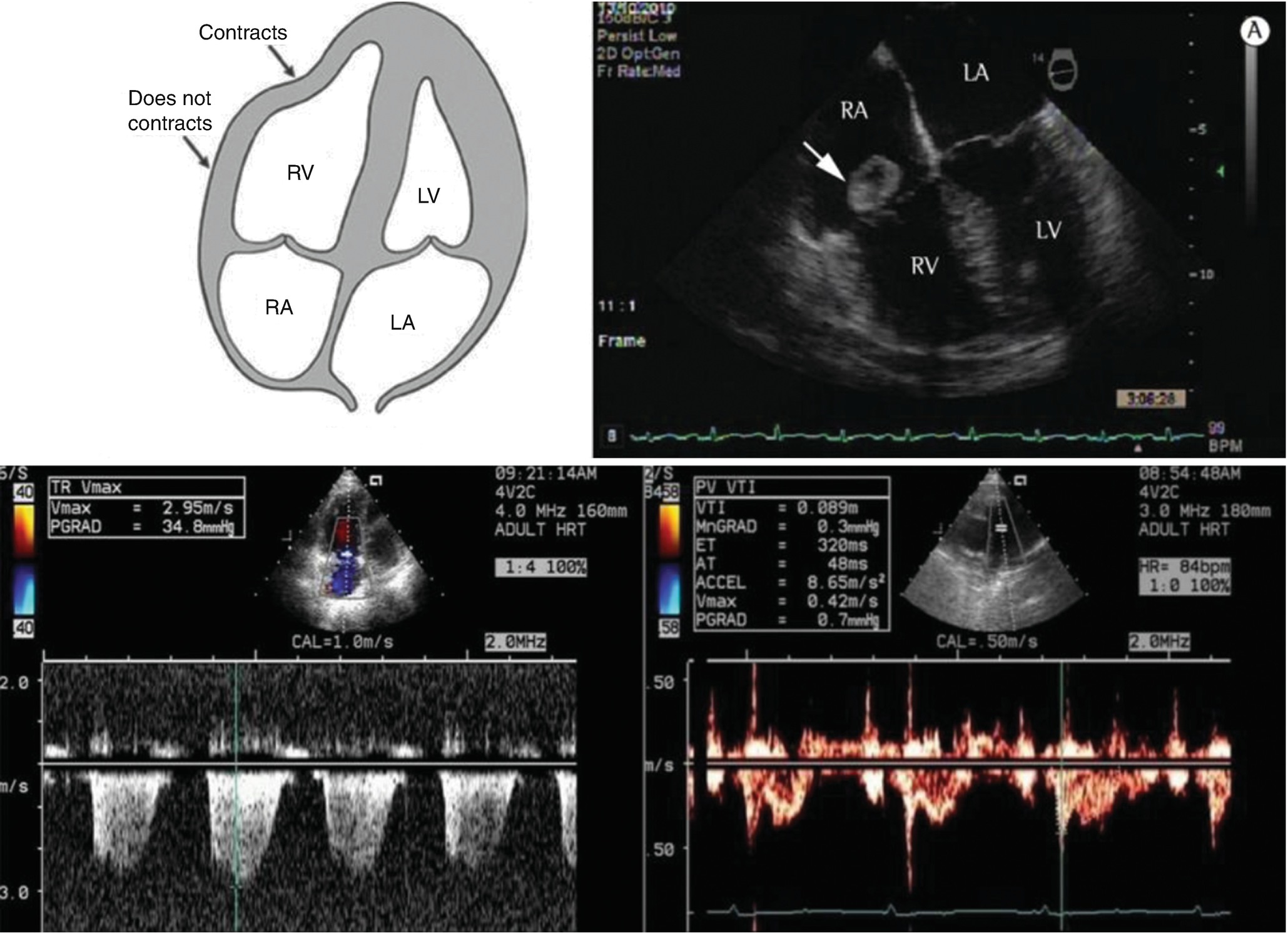


Echocardiographic Evaluation Of The Right Heart Springerlink
Normal values for aorta in 2D echocardiography Normal interval Normal interval, adjusted Aortic annulus 31 mm 1214 mm/m2 Sinus valsalva 2945 mm 15 mm/m2A Report from the American Society of Echocardiography Developed in Collaboration with the Society for Cardiovascular Magnetic Resonance In Journal of the American Society of Echocardiography official publication of the American Society of Echocardiography 30 (4), pp 303–371 DOI /jecho7 –>PubmedLink 3 Normal values for Dopplerderived diastolic measurements Age group (y) Measurement 16 2140 4160 >60 IVRT (ms) 50 ± 9 (3268) 67 ± 8 (51) 74 ± 7 (60) 87 ± 7 () E/A ratio 1 ± 045 (0978) 153 ± 040 () 128 ± 025 () 096 ± 018 () DT (ms) 142 ± 19 () 166 ± 14 ()


Global And Regional Right Ventricular Function Assessed By Novel Three Dimensional Speckle Tracking Echocardiography Journal Of The American Society Of Echocardiography
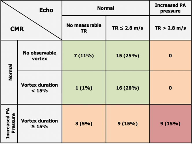


Cardiovascular Magnetic Resonance 4d Flow Analysis Has A Higher Diagnostic Yield Than Doppler Echocardiography For Detecting Inc
What is a transthoracic echocardiogram (TTE)? The normal values of left and right ventricle differ Due to the high pressures which must be generated by the LV, the isovolumic times of the LV are relatively long The RV generates less pressure and therefore the index also lower with an average at around 028 A Tei index above 055 (TDI) is considered abnormalECHO or TRPG for detecting a mean PAP 25 mmHg that was determined invasively has not been well defined We studied 1 patients who underwent RHC Echocardiography was performed within 24 hours of invasive evaluation, and sPAP ECHO was defined as the TRPG with right atrial pressure estimated on the basis of the current guideline


Updated Left Ventricular Diastolic Function Recommendations And Cardiovascular Events In Patients With Heart Failure Hospitalization Journal Of The American Society Of Echocardiography


Csbosysgnewfym
The Doppler definition of PAH is based on tricuspid regurgitation (TR) jet that is > 28 m/sec Utilizing RAP estimation of 5 to 15 mm Hg (most commonly used measurements depending on the characteristics of IVC), this equals SPAP of 36 mm Hg to 46 mm Hg


Advanced Imaging In Pulmonary Hypertension Thoracic Key


Exaggerated Increase Of Exercise Induced Pulmonary Artery Pressure In Systemic Sclerosis Patients Predominantly Results From Left Ventricular Diastolic Dysfunction Springerlink


Echo Test Results Normal


Jvs Journal Of Veterinary Science


Echocardiographic Assessment Of The Right Ventricle State Of The Art Sciencedirect


Emdocs Net Emergency Medicine Educationus Probe Tapse Emdocs Net Emergency Medicine Education


Pulmonary Hypertension


A Subclinical High Tricuspid Regurgitation Pressure Gradient Independent Of The Mean Pulmonary Artery Pressure Is A Risk Factor For The Survival After Living Donor Liver Transplantation Bmc Gastroenterology Full Text


A Decrease In Tricuspid Regurgitation Pressure Gradient Associates With Favorable Outcome In Patients With Heart Failure Seko Esc Heart Failure Wiley Online Library


Www Asecho Org Wp Content Uploads 18 01 Kusunose Pulmonary Hypertension And Pulmonary Embolism Role Of Echo Pdf


Diagnostic Value Of Right Pulmonary Artery Distensibility Index In Dogs With Pulmonary Hypertension Comparison With Doppler Echocardiographic Estimates Of Pulmonary Arterial Pressure Abstract Europe Pmc


Tricuspid Regurgitation Peak Gradient Trpg Tricuspid Annulus Plane Systolic Excursion Tapse A Novel Parameter For Stepwise Echocardiographic Risk Stratification In Normotensive Patients With Acute Pulmonary Embolism Semantic Scholar


6 Tips For Calculating Rvsp


Pulmonary Hypertension


Effusions Explored


Tricuspid Annular Plane Systolic Excursion Tapse For Risk Stratification And Prognostication Of Patients With Pulmonary Embolism Journal Of Emergency Medicine


Overestimation By Echocardiography Of The Peak Systolic Pressure Gradient Between The Right Ventricle And Right Atrium Due To Tricuspid Regurgitation And The Usefulness Of The Early Diastolic Transpulmonary Valve Pressure Gradient For


Www Heartlungcirc Org Article S1443 9506 19 X Pdf


Csbosysgnewfym


Prognostic Significance Of Right Ventricular Function During Exercise In Asymptomatic Minimally Symptomatic Patients With Nonobstructive Hypertrophic Cardiomyopathy Hirasawa Echocardiography Wiley Online Library


Cardiovascular Magnetic Resonance 4d Flow Analysis Has A Higher Diagnostic Yield Than Doppler Echocardiography For Detecting Increased Pulmonary Artery Pressure Springerlink


Web Downloadable Software For Training In Cardiovascular Hemodynamics In The 3 D Stress Echo Lab Topic Of Research Paper In Medical Engineering Download Scholarly Article Pdf And Read For Free On Cyberleninka


Aortic Sclerosis Diagnosis Treatments Risk Factors


Tricuspid Regurgitation Peak Gradient Trpg Tricuspid Annulus Plane Systolic Excursion Tapse A Novel Parameter For Stepwise Echocardiographic Risk Stratification In Normotensive Patients With Acute Pulmonary Embolism Semantic Scholar


2


Www Researchsquare Com Article Rs V1 Pdf


Diagnostic Value Of Right Pulmonary Artery Distensibility Index In Dogs With Pulmonary Hypertension Comparison With Doppler Echocardiographic Estimates Of Pulmonary Arterial Pressure Visser 16 Journal Of Veterinary Internal Medicine Wiley


Qualification Of Patients For Right Heart Catheterization Tte Download Scientific Diagram


Cardiovascular Magnetic Resonance 4d Flow Analysis Has A Higher Diagnostic Yield Than Doppler Echocardiography For Detecting Increased Pulmonary Artery Pressure Springerlink


Carnitine Insufficiency Is Associated With Adverse Outcomes In Patients With Heart Failure With Preserved Ejection Fraction Jarlife


2


16 2 2 How To Assess Pulmonary Hypertension 123 Sonography


Jvs Journal Of Veterinary Science


Characteristics And Prognostic Associations Of Echocardiographic Pulmonary Hypertension With Normal Left Ventricular Systolic Function In Patients 90 Years Of Age Sciencedirect


Jvs Journal Of Veterinary Science


2


Exaggerated Increase Of Exercise Induced Pulmonary Artery Pressure In Systemic Sclerosis Patients Predominantly Results From Left Ventricular Diastolic Dysfunction Topic Of Research Paper In Clinical Medicine Download Scholarly Article Pdf And Read


Comprehensive Cardiovascular Magnetic Resonance Diastolic Dysfunction Grading Shows Very Good Agreement Compared With Echocardiography Jacc Cardiovascular Imaging


18 Tsoc Guideline Focused Update On Diagnosis And Treatment Of Pulmonary Arterial Hypertension Sciencedirect
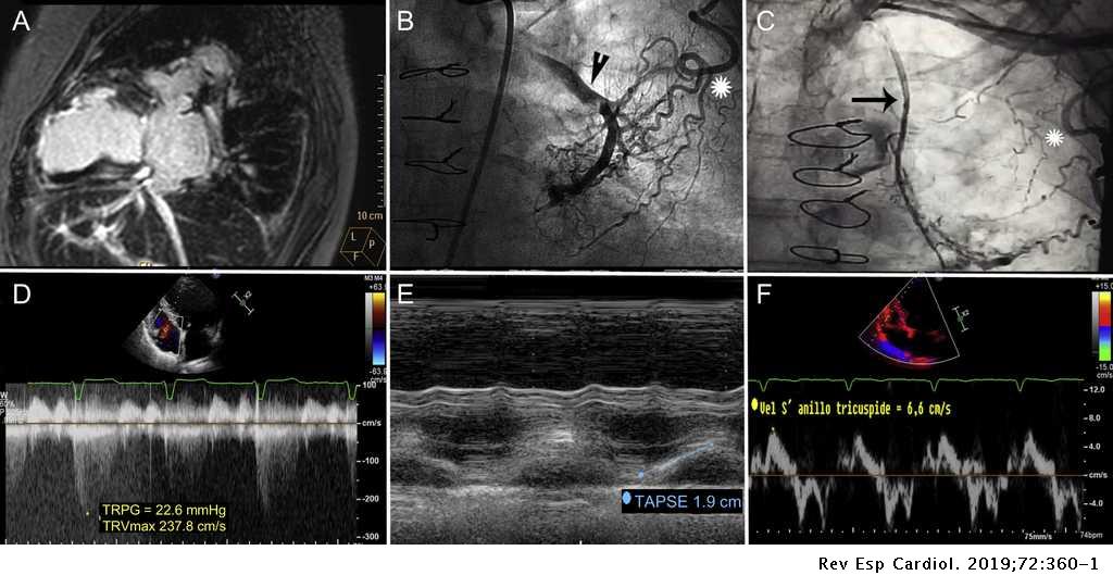


Percutaneous Venous Pulmonary Artery Extracorporeal Membrane Oxygenation In Right Heart Failure Revista Espanola De Cardiologia


Http Repository Tnmgrmu Ac In 6614 1 lathapk Pdf


Pdf Tricuspid Regurgitation Peak Gradient Trpg Tricuspid Annulus Plane Systolic Excursion Tapse A Novel Parameter For Stepwise Echocardiographic Risk Stratification In Normotensive Patients With Acute Pulmonary Embolism


Figure 2 From Assessment Of The American Society Of Echocardiography European Association Of Echocardiography Guidelines For Diastolic Function In Patients With Depressed Ejection Fraction An Echocardiographic And Invasive Haemodynamic Study


How To Estimate Pulmonary Artery Systolic Pressure Pasp Using Echo Youtube


Extravascular Lung Water And Cardiac Function Assessed By Echocardiography In Healthy Lowlanders During Repeated Very High Altitude Exposure International Journal Of Cardiology


Comprehensive Cardiovascular Magnetic Resonance Diastolic Dysfunction Grading Shows Very Good Agreement Compared With Echocardiography Jacc Cardiovascular Imaging


Diagnostic Value Of Right Pulmonary Artery Distensibility Index In Dogs With Pulmonary Hypertension Comparison With Doppler Echocardiographic Estimates Of Pulmonary Arterial Pressure Abstract Europe Pmc


Linear Regression Solid Line Of Estimated Mean Pulmonary Artery Download Scientific Diagram


Echocardiography Monitoring Of Pulmonary Hypertension After Pediatric Hematopoietic Stem Cell Transplantation Pediatric Pulmonary Arterial Hypertension And Pulmonary Veno Occlusive Disease After Hematopoietic Stem Cell Transplantation
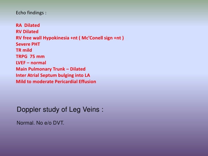


A Case Of Acute Rvf


Pdf Echocardiographic Evaluation Of The Right Atrial Area Index In Dogs With Pulmonary Hypertension


2


Rv Doppler Resistance Vs Back Pressure Jon Emile Kenny Korbin Haycock Foamed Foamer Foamcc Pocus Thinking Critical Care


1
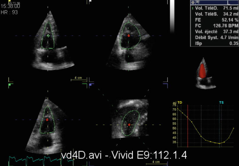


Advanced Imaging In Pulmonary Hypertension Thoracic Key


Left Atrial Strain Associated With Functional Recovery In Patients Receiving Optimal Treatment For Heart Failure Journal Of The American Society Of Echocardiography


Abstract Echocardiography Wiley Online Library


Right Ventricular Dyssynchrony Predicts Clinical Outcomes In Patients With Pulmonary Hypertension International Journal Of Cardiology
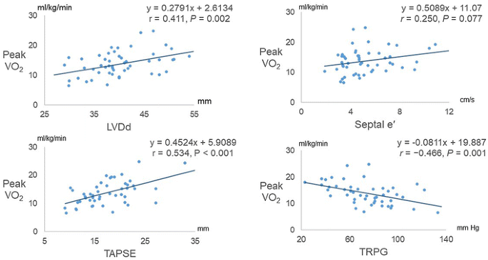


Left Ventricular End Diastolic Dimension And Septal E Are Predictors Of Cardiac Index At Rest While Tricuspid Annular Plane Systolic Excursion Is A Predictor Of Peak Oxygen Uptake In Patients With Pulmonary Hypertension


Diagnostic Value Of Right Pulmonary Artery Distensibility Index In Dogs With Pulmonary Hypertension Comparison With Doppler Echocardiographic Estimates Of Pulmonary Arterial Pressure Visser 16 Journal Of Veterinary Internal Medicine Wiley


Cdn Doctorsonly Co Il 18 06 A Puzzling Case Of Pulmonary Hypertension Dr Michal Gur Pdf
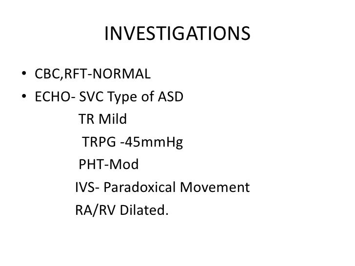


A Case Of Asd Sinus Venosus Type


Echocardiography Monitoring Of Pulmonary Hypertension After Pediatric Hematopoietic Stem Cell Transplantation Pediatric Pulmonary Arterial Hypertension And Pulmonary Veno Occlusive Disease After Hematopoietic Stem Cell Transplantation


Openventio Org Wp Content Uploads 17 10 Serial Measurements Of Tricuspid Regurgitation Pressure Gradient By Echocardiography Predict Prognosis In Idiopathic Pulmonary Fibrosis Prrmoj 3 124 Pdf


16 2 2 How To Assess Pulmonary Hypertension 123 Sonography
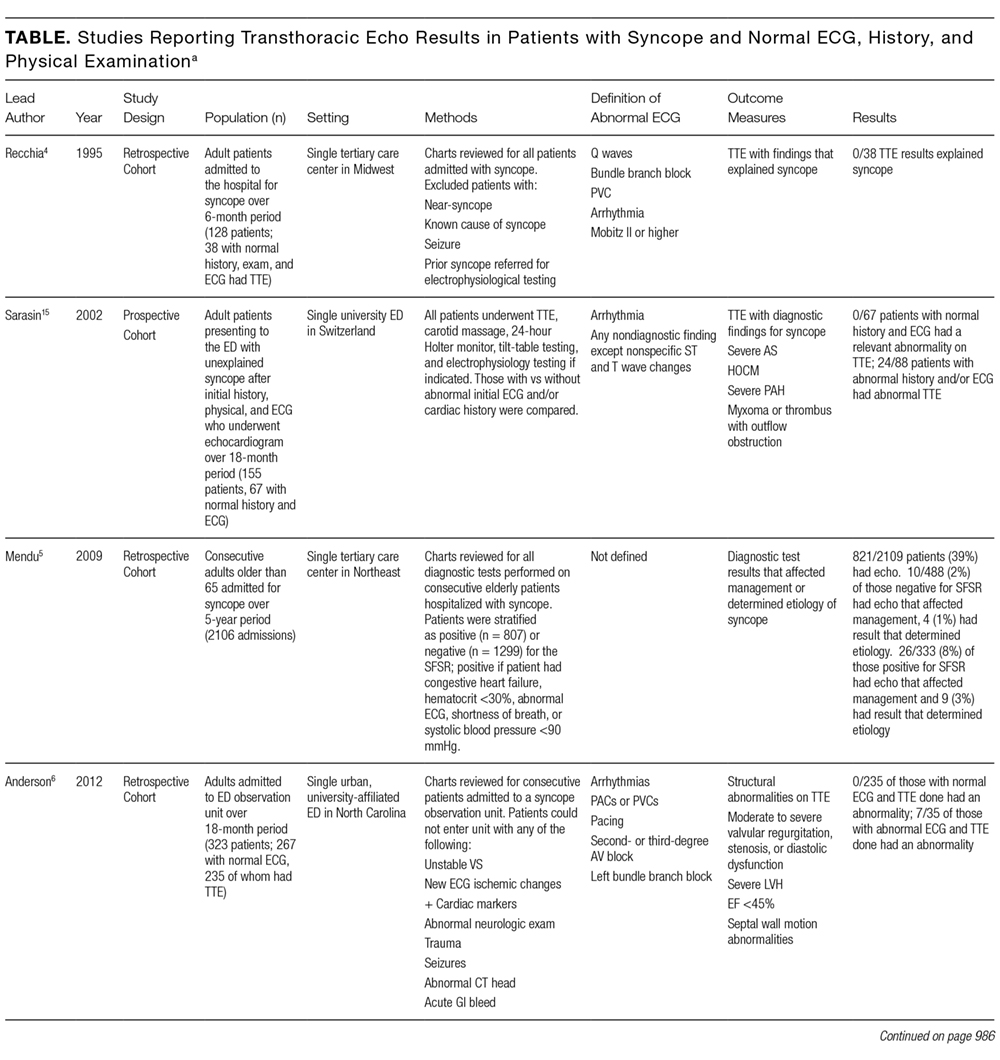


Echo Test Results Normal
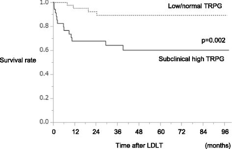


A Subclinical High Tricuspid Regurgitation Pressure Gradient Independent Of The Mean Pulmonary Artery Pressure Is A Risk Factor For The Survival After Living Donor Liver Transplantation Bmc Gastroenterology Full Text


Abstract Echocardiography Wiley Online Library


Cardiovascular Magnetic Resonance 4d Flow Analysis Has A Higher Diagnostic Yield Than Doppler Echocardiography For Detecting Increased Pulmonary Artery Pressure Research Square
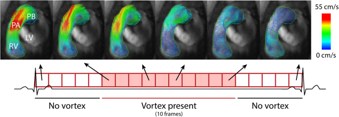


Cardiovascular Magnetic Resonance 4d Flow Analysis Has A Higher Diagnostic Yield Than Doppler Echocardiography For Detecting Increased Pulmonary Artery Pressure Springerlink


A Case Of Acute Rvf


Q Tbn And9gcredsygn7e38r3z2rolqxin85lgeed8enm8evvr7hs4v0o2lhtq Usqp Cau


Echocardiography Of Chronic Right Heart Failure Thoracic Key


Pvrinstitute Org Media 62 Diagnostic Algorithm V3 Pdf


Cdn Doctorsonly Co Il 18 06 A Puzzling Case Of Pulmonary Hypertension Dr Michal Gur Pdf


Jvs Journal Of Veterinary Science


Echo Test Results Normal


开放获取论文一站式发现平台


Pdf Tricuspid Regurgitation Peak Gradient Trpg Tricuspid Annulus Plane Systolic Excursion Tapse A Novel Parameter For Stepwise Echocardiographic Risk Stratification In Normotensive Patients With Acute Pulmonary Embolism


Left Ventricular End Diastolic Dimension And Septal E Are Predictors Of Cardiac Index At Rest While Tricuspid Annular Plane Systolic Excursion Is A Predictor Of Peak Oxygen Uptake In Patients With Pulmonary Hypertension


0 件のコメント:
コメントを投稿